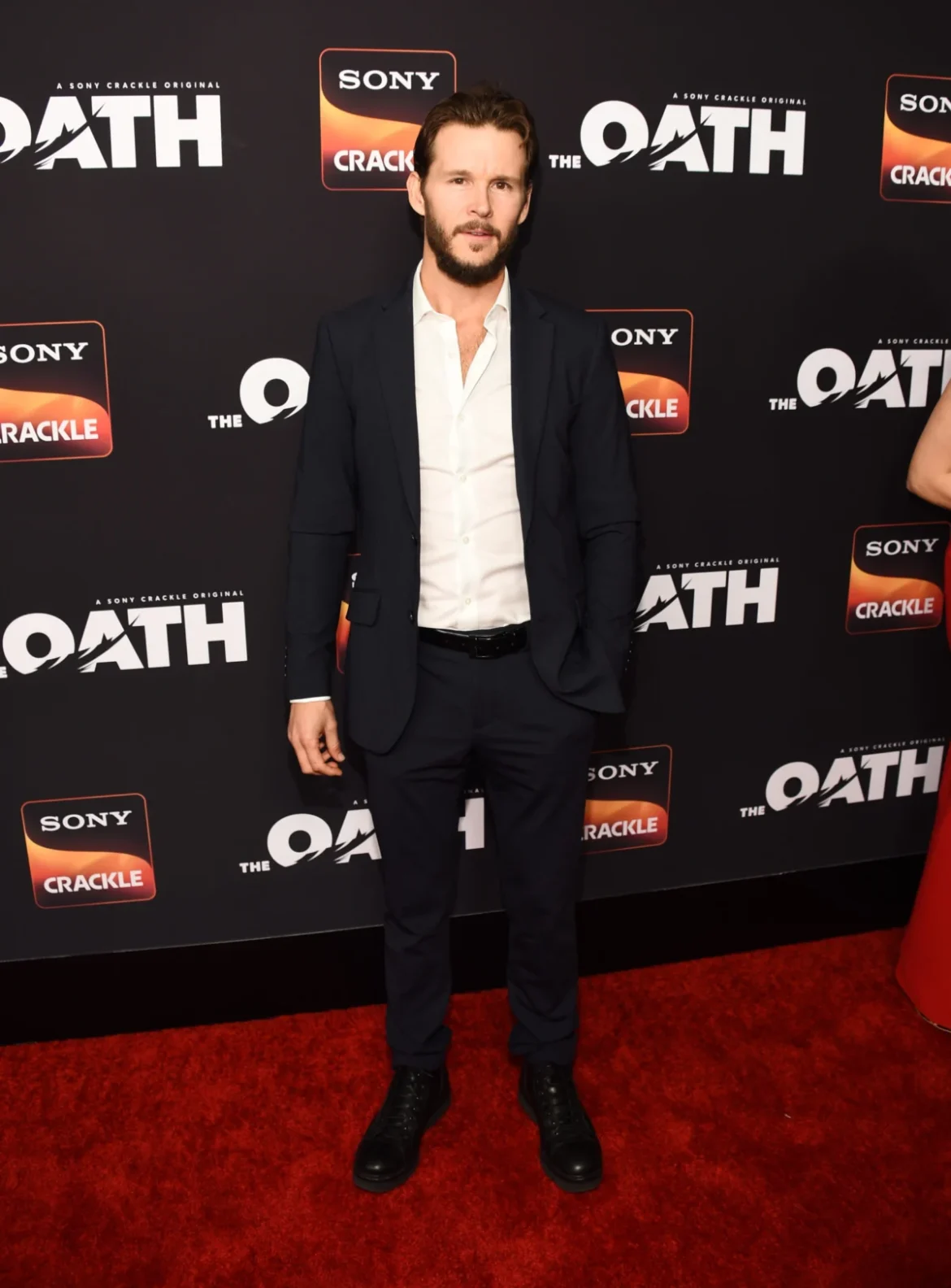
The landscape of talent management in the entertainment industry continues to evolve as established artists align themselves with forward-thinking firms to navigate an increasingly complex media ecosystem. In the latest development signaling this ongoing shift, veteran Australian actor and producer Ryan Kwanten has officially signed with Rain for exclusive management representation. The strategic alliance places Kwanten within a robust stable of creative talent managed by the firm, positioning the multifaceted performer for the next phase of a career that spans over three decades across television, film, and digital platforms.
Kwanten, whose career began in the early 1990s in his native Australia before expanding into the competitive U.S. market, maintains representation with RGM Artists for his domestic and international endeavors. The addition of Rain to his team marks a pivotal moment as the actor looks to broaden his footprint in both starring roles and behind-the-camera production ventures, leveraging the firm’s industry relationships to secure high-profile projects in an era dominated by streaming platforms and global content demand.
Early Beginnings and the Australian Television Foundation
The foundational years of Ryan Kwanten’s career were established in the rigorous environment of Australian television production during the golden era of local serial dramas. Born in Sydney, New South Wales, Kwanten demonstrated an early aptitude for performance, securing his first professional acting credits in the early 1990s. His steady progression through minor guest roles on popular series laid the groundwork for his eventual breakthrough.
The definitive turning point of his early career arrived when he cast in the iconic Australian soap opera Home and Away. Airing on the Seven Network, the long-running serial has famously served as a launching pad for numerous international stars. Kwanten’s tenure on the program allowed him to hone his craft under the demanding daily production schedules typical of daytime television, building a dedicated fan base and establishing a reputation for reliability and screen presence.
Capitalizing on the international reach of Australian television talent, Kwanten successfully transitioned to the American market in the mid-2000s. He secured a prominent role in the WB network drama series Summerland, created by Stephen Tolkin and Lori Loughlin. Playing Jay Robertson, Kwanten charmed audiences and demonstrated his ability to adapt to the sensibilities of American prime-time television, setting the stage for the career-defining role that would soon follow on premium cable.
The True Blood Phenomenon and Global Recognition
In 2008, Kwanten achieved international stardom when he was cast in Alan Ball’s groundbreaking HBO fantasy-horror series True Blood. Adapted from Charlaine Harris’s bestselling book series The Southern Vampire Mysteries, the show became a massive cultural and commercial phenomenon for the network.
Kwanten portrayed Jason Stackhouse, the perpetually bewildered yet fiercely loyal and endearing brother of telepathic waitress Sookie Stackhouse (played by Anna Paquin). Across the series’ eight-season run, which concluded in 2014 after 81 episodes, Kwanten’s portrayal of Jason evolved from a superficial small-town heartthrob into a complex, morally conflicted character grappling with the supernatural realities of Bon Temps, Louisiana.
True Blood cemented HBO’s dominance in the Sunday night prestige television slot and earned widespread critical acclaim, including a Primetime Emmy Award nomination and multiple Saturn Awards. For Kwanten, the series provided a global platform, showcasing his comedic timing, physical acting capabilities, and dramatic range to casting directors and producers worldwide. The role established him as a recognizable face in households across the globe and opened doors to diverse cinematic opportunities.
Diversifying the Portfolio: Film Credits and Executive Producing
Following the conclusion of True Blood, Kwanten deliberately cultivated a diverse filmography that defied typecasting, traversing independent cinema, genre thrillers, and big-budget studio projects. His cinematic endeavors span a wide array of genres, working alongside some of the most respected actors and directors in the global film industry.
In the psychological horror-thriller Glorious (2022), directed by Rebekah McKendry, Kwanten delivered a tour-de-force performance opposite veteran actor J.K. Simmons. Beyond his starring duties, Kwanten took on the role of executive producer, signaling his growing interest in the creative and financial orchestration of independent filmmaking. His hands-on approach to production demonstrated a sophisticated understanding of independent film financing, packaging, and distribution strategies.
Kwanten’s versatility is further underscored by his collaborations with acclaimed international auteurs and legendary screen actors. He starred alongside Academy Award winner Nicolas Cage in the geopolitical crime thriller Kill Chain (2019), directed by Ken Sanzel. He also shared the screen with Academy Award winner Sir Anthony Hopkins in the biographical crime film Kidnapping Mr. Heineken (2015), directed by Daniel Alfredson, which chronicled the sensational 1983 abduction of the Dutch brewing magnate Freddy Heineken.
In the realm of voice acting and animation, Kwanten lent his vocal talents to Zack Snyder’s critically acclaimed fantasy adventure Legend of the Guardians: The Owls of Ga’Hoole (2010). Voicing the character of Kludd, Kwanten joined an elite ensemble cast that included Helen Mirren, Abbie Cornish, Joel Edgerton, Sam Neill, Hugo Weaving, Jim Sturgess, and Geoffrey Rush. The project highlighted his ability to deliver nuanced performances without the benefit of physical onscreen presence.
Recent Ventures and Upcoming Projects
In recent years, Kwanten has maintained an aggressive production schedule, balancing television series regular roles with independent feature films. In 2018, he starred in and executive-produced the Crackle crime drama series The Oath, collaborating with executive producer Curtis “50 Cent” Jackson. The gritty police procedural explored the complex subculture of corrupt gangs within law enforcement agencies.
His television credits continued to expand with appearances in Amazon Prime Video’s acclaimed anthology horror series Them, as well as roles in Kindred, Head Count, and the horror-mystery feature The Portrait (2023). Demonstrating his affinity for historical and action-oriented narratives, Kwanten recently completed work on the World War II-set combat film Primitive War and the military action thriller Seven Snipers, alongside seasoned cinematic veterans Tim Roth, Ioan Gruffudd, and Radha Mitchell.
Looking ahead, Kwanten is slated to star in the upcoming post-apocalyptic werewolf thriller Dead Howling, a project that promises to further cement his status as a bankable genre lead. His consistent output across multiple mediums illustrates a calculated career trajectory focused on longevity, creative control, and active participation in project development.
Strategic Implications of the Rain Partnership
The management partnership with Rain arrives at a crucial juncture in the entertainment industry. In the wake of industry contractions, strikes, and shifting models of content commissioning, talent management firms are tasked with identifying alternative funding sources, digital-first opportunities, and international co-productions for their clients.
By securing representation with Rain, Kwanten is positioned to leverage the firm’s specialized expertise in packaging and strategic career planning. Rain represents a roster of dynamic creators, writers, directors, and actors, fostering an environment of collaborative synergy. For Kwanten, who has increasingly stepped behind the camera as an executive producer, the firm’s capabilities in independent financing and development will be instrumental in bringing proprietary intellectual property to the screen.
Industry analysts note that veteran actors who successfully pivot into producing roles—much like Kwanten has done with projects such as Glorious and The Oath—enjoy greater career sustainability. By managing both their on-screen presence and behind-the-scenes stakes, artists can insulate themselves from the cyclical nature of acting employment. Rain’s intervention is expected to accelerate Kwanten’s ambitions in the producing sphere while strategically curating his next wave of acting assignments across studio and independent platforms.
Future Outlook for Ryan Kwanten
As the entertainment landscape pivots toward globalized streaming metrics and diversified content consumption, the demand for adaptable, globally recognized talent remains high. Ryan Kwanten’s extensive résumé, stretching from Australian serial television to prestige American cable and international independent cinema, provides a solid foundation for continued relevance.
With his ongoing representation by RGM Artists ensuring a strong domestic and international bridge, and Rain now steering his broader career strategy, Kwanten enters the next chapter of his professional life with a fortified support network. As production on projects like Dead Howling wraps and new development ventures take shape, the actor-producer remains a compelling figure to watch in contemporary film and television.