
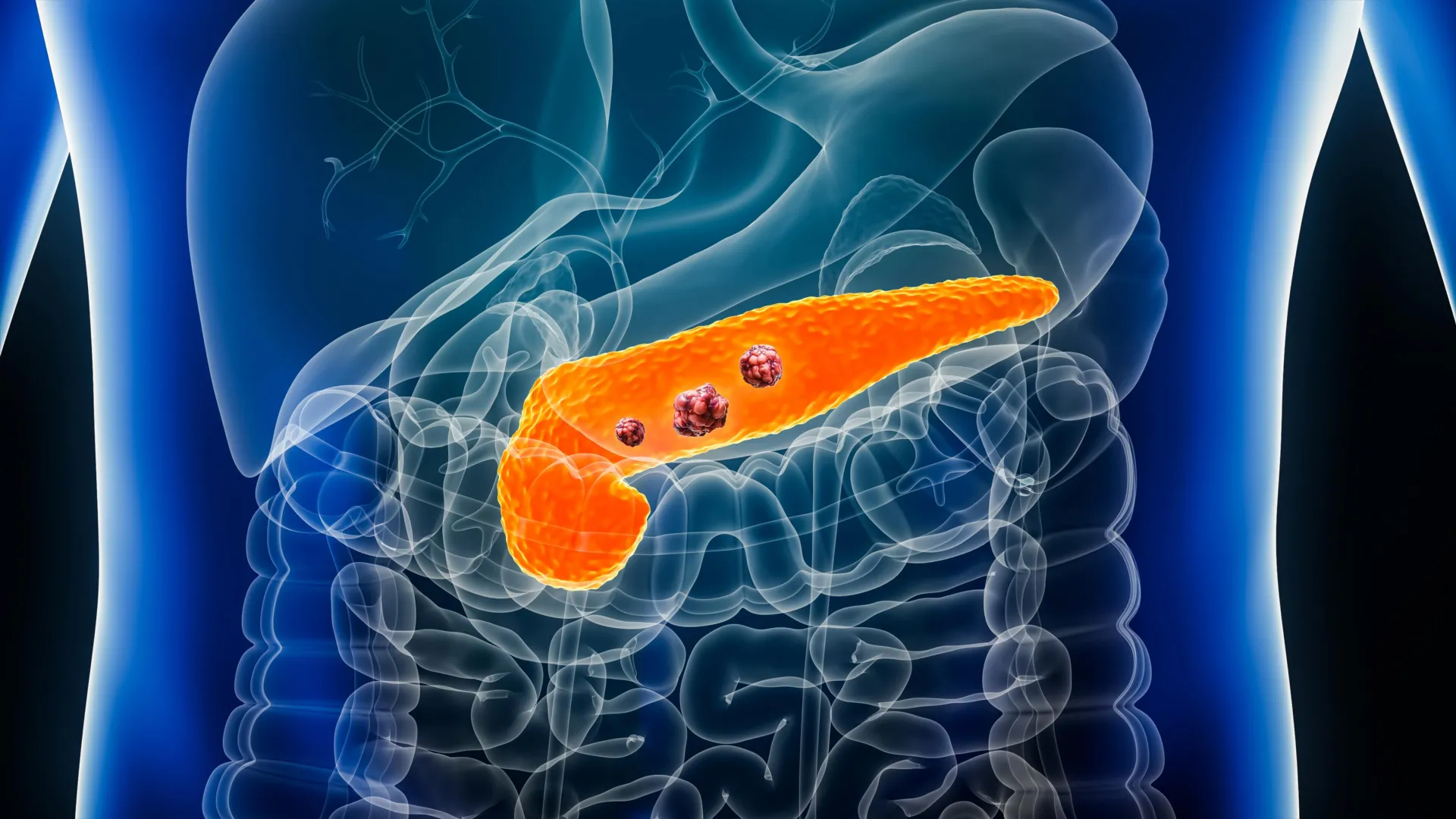
Pancreatic ductal adenocarcinoma remains one of the most formidable adversaries in modern oncology, primarily because it is frequently diagnosed only after the disease has advanced to a terminal stage. However, a new artificial intelligence framework known as REDMOD (Radiomics-based Early Detection MODel) is poised to alter this clinical reality. Published online in the medical journal Gut, a recent multi-center study demonstrates that REDMOD can identify extremely subtle tissue alterations indicative of early-stage pancreatic cancer long before conventional imaging technologies or expert human eyes can perceive them. By analyzing microscopic texture variations within standard abdominal computed tomography (CT) scans, the AI system successfully detected hidden cancer signatures an average of 475 days prior to clinical diagnosis. This technological leap offers a prospective window for early intervention, potentially shifting the paradigm of pancreatic cancer management from reactive late-stage treatment to proactive, pre-clinical interception.
The Anatomy of a Silent Killer: Why Early Detection Fails
To understand the magnitude of the advancement represented by REDMOD, one must first examine the inherent challenges of diagnosing pancreatic ductal adenocarcinoma (PDAC). The pancreas is a vital, deeply seated organ located behind the stomach, making it difficult to visualize clearly through routine physical examinations. Furthermore, early-stage pancreatic cancer is notoriously asymptomatic. When symptoms do finally manifest—such as jaundice, abdominal pain, or unexplained weight loss—the disease has typically metastasized or invaded surrounding major blood vessels, drastically limiting treatment options.
Standard diagnostic pathways rely heavily on conventional cross-sectional imaging, most notably computed tomography (CT) scans. Yet, the early microscopic changes that occur within the pancreatic parenchyma often produce visual alterations that blend seamlessly with normal tissue to the human eye. Radiologists, despite years of rigorous training and extensive clinical experience, struggle to differentiate these subtle textural shifts from benign variations. Consequently, the five-year survival rate for pancreatic cancer remains critically low compared to many other malignancies. Medical experts have long maintained that the single most critical determinant of survival is the timing of diagnosis. Statistical modeling underscores this clinical reality: increasing the proportion of localized pancreatic cancers detected at stage 0 from the current baseline of approximately 10% to 50% would more than double overall survival rates.
Chronology of the Study: Tracing Scans Back in Time
To rigorously test the efficacy of the REDMOD framework, an international team of researchers designed a retrospective, multi-center study that analyzed historical medical records and abdominal CT scans. The primary objective was to determine whether artificial intelligence could identify predictive imaging signatures in scans previously deemed normal by expert radiologists.
The research cohort comprised 219 patients who were treated across multiple medical institutions and subsequently diagnosed with pancreatic ductal adenocarcinoma. Crucially, the abdominal CT scans evaluated in the study had been performed months or even years prior to their actual clinical diagnosis, at which point experienced radiologists had documented no evidence of malignancy.
The researchers stratified these historical scans into distinct temporal intervals to gauge how far in advance the AI could peer into the pre-diagnostic window:
- 40% of the patients (87 individuals) had undergone CT imaging between 3 and 12 months prior to their clinical diagnosis.
- 35% of the patients (76 individuals) had scans taken 12 to 24 months before diagnosis.
- 25% of the patients (56 individuals) had imaging performed more than 24 months prior to diagnosis, with some timelines stretching back to approximately three years.
Anatomically, the disease distribution within this cohort aligned with epidemiological norms for the malignancy, with nearly two-thirds (64%) of the tumors originating in the head of the pancreas. The patients who eventually developed the disease had an average age of 69 years, spanning a wide age range from 34 to 88 years.
To establish a robust control group, the scientists gathered CT scans from 1,243 individuals who remained entirely free of pancreatic cancer for up to three years following their imaging sessions. This comparison cohort was meticulously matched to the cancer group based on age, sex, and scan dates, yielding an average age of 64 years within the same 34-to-88 age span.
Quantitative Performance: AI Surpasses Human Expertise
When the REDMOD framework was deployed across this extensive dataset, its diagnostic performance significantly outstripped that of human specialists. The comparative metrics underscore a profound divergence in capability between machine learning algorithms and traditional radiological assessment.
In evaluating sensitivity—the statistical measure of how effectively a diagnostic tool correctly identifies individuals who genuinely possess a disease—REDMOD achieved an overall sensitivity of 73%. In stark contrast, the experienced radiologists who originally reviewed or re-evaluated the same historical scans achieved a sensitivity of just 39%.
The performance gap widened even further when researchers analyzed the subset of scans captured more than two years before clinical detection. In these exceptionally challenging cases, where the disease was in its absolute infancy, REDMOD maintained a detection rate of 68%. Human radiologists, conversely, recorded a detection rate of only 23% for the exact same set of long-lead-time images, meaning the AI performed nearly three times more effectively at spotting deep historical signals.
Specificity and Reliability Testing
Beyond its ability to catch hidden malignancies, a clinically viable diagnostic tool must also demonstrate high specificity to minimize false positives and avoid subjecting healthy patients to unnecessary invasive procedures. To test this, the researchers evaluated REDMOD on an independent validation cohort of 539 patients from several distinct hospital systems who did not have pancreatic cancer. The AI successfully classified just over 81% of these scans as definitively cancer-free.
To further validate these findings against external benchmarks, the model was tested on a publicly available dataset from the United States National Institutes of Health (the NIH-PCT dataset, comprising 80 patients). On this standardized external data, REDMOD correctly identified 87.5% of the scans as free of pancreatic cancer, proving its robustness across different imaging protocols and hardware manufacturers.
Consistency is another vital benchmark for automated medical diagnostics. To determine whether REDMOD’s early warning signs were stable over time, researchers analyzed multiple scans taken from the same patients several months apart. The system produced matching, consistent classifications in 90% to 92% of these sequential cases, providing strong evidence that the algorithm was locking onto genuine, persistent biological changes rather than transient imaging artifacts or scan noise.
Technological Innovation: Radiomics and Automated Segmentation
The underlying engine driving REDMOD’s success is the science of radiomics—the high-throughput extraction of large amounts of quantitative features from medical images. While the human visual cortex is limited to perceiving gross macroscopic shapes, density variations, and obvious structural distortions, radiomics algorithms can quantify microscopic spatial pixel-intensity variations, texture heterogeneity, and edge gradients within an organ’s tissue. In the case of pancreatic ductal adenocarcinoma, the tumor and its surrounding microenvironment induce minute alterations in tissue architecture that alter how X-rays pass through the organ during a CT scan. REDMOD’s algorithms are specifically tuned to recognize these invisible textural signatures.
Furthermore, the framework addresses a notorious bottleneck in clinical workflow: organ segmentation. Traditionally, evaluating an abdominal CT scan requires a radiologist or specialized technician to manually outline the boundaries of the pancreas slice by slice. This manual process is not only time-consuming but also introduces inter-observer variability, as different clinicians may trace slightly different boundaries. REDMOD automates this entire process by instantly and accurately identifying, isolating, and outlining the pancreas, separating it cleanly from adjacent structures such as the duodenum, spleen, and major vascular networks. By standardizing boundary delineation, the AI eliminates human inconsistency and ensures that the subsequent radiomic analysis is performed on the exact same anatomical volume every time.
Broader Clinical Implications and Expert Analysis
The publication of the REDMOD study has generated considerable discussion within the global oncology and radiology communities. Medical analysts point out that if prospective trials confirm these retrospective findings, the implications for healthcare systems could be transformative. Pancreatic cancer care currently consumes disproportionate healthcare resources due to the intensive costs associated with late-stage palliative therapies, chemotherapy resistance, and complex surgeries that ultimately arrive too late.
Shifting the detection timeline upstream by an average of 475 days means that patients could be identified at stage 0. At this pre-invasive or early invasive stage, surgical resection—currently the only potential cure for pancreatic cancer—carries a vastly higher success rate. Moreover, patients would be spared the physical degradation of advanced disease and could potentially undergo less aggressive, more targeted therapeutic regimens.
Despite the enthusiasm surrounding these findings, the study’s authors and independent clinical commentators emphasize that several critical hurdles remain before REDMOD can be integrated into routine hospital workflows. Foremost among these is the need for rigorous prospective clinical trials. Retrospective studies, while powerful for generating hypotheses and proving retrospective accuracy, must be validated in real-time prospective settings where the AI evaluates incoming scans from living patients as they present to clinics.
Additionally, the researchers noted limitations regarding the demographic diversity of the current study population. The training and validation cohorts did not include a sufficiently diverse representation of various ethnic and racial backgrounds. Because anatomical variations, body mass indexes, and baseline tissue densities can differ across populations, the algorithm must be tested and retrained across globally diverse cohorts to ensure equitable performance and to prevent algorithmic bias.
Next Steps for Clinical Integration
Looking ahead, clinical researchers are planning targeted prospective studies focusing on high-risk patient populations. Rather than deploying the tool indiscriminately across the general public, initial clinical implementation will likely target individuals exhibiting known risk factors for pancreatic cancer. These high-risk groups include patients presenting with new-onset diabetes later in life, individuals suffering from unexplained and rapid weight loss, those with a strong familial history of pancreatic malignancies, or patients diagnosed with chronic pancreatitis.
As machine learning continues to mature within medical imaging departments, frameworks like REDMOD signal a profound shift in how physicians interact with diagnostic data. Rather than replacing the radiologist, tools of this caliber are designed to function as an advanced early-warning radar system, flagging high-risk scans that demand closer human inspection or follow-up imaging.
The research team concluded their study by emphasizing the broader mission of their work: "While prospective validation is paramount to confirm clinical utility, the REDMOD framework represents a significant advance towards shifting the paradigm for sporadic pancreatic ductal adenocarcinoma from a late-stage symptomatic diagnosis to proactive pre-clinical interception, offering tangible hope for improving outcomes in this challenging disease." As validation trials progress over the coming years, the medical community moves steadily closer to turning a once-invisible diagnosis into a manageable, treatable condition.

