
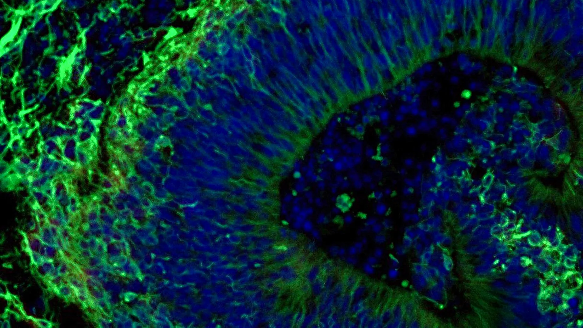
Scientists at Johns Hopkins Medicine have identified groundbreaking new evidence suggesting that miniature brain tissues, cultivated from the cells of individuals with Alzheimer’s disease, could serve as a powerful tool for predicting how different patients might respond to medications designed to manage the condition’s associated psychiatric symptoms. This innovative research, focusing on laboratory-grown brain organoids, adds significant weight to the growing body of evidence supporting the use of these sophisticated models in developing and selecting more precise and individualized treatment strategies for Alzheimer’s disease, a progressive neurodegenerative disorder that stands as the most common form of dementia, impacting an estimated 7 million Americans.
The implications of this study extend beyond personalized treatment selection. Researchers also discovered that these organoids release minuscule particles known as extracellular vesicles, which act as carriers of vital cellular information. These vesicles hold considerable promise as potential biomarkers for both the early diagnosis of Alzheimer’s disease and the accurate assessment of its progression. The findings, which received partial funding from the National Institutes of Health (NIH), have been formally published in the esteemed journal Alzheimer’s & Dementia: The Journal of the Alzheimer’s Association.
Mini Brain Models Pave the Way for Precision Alzheimer’s Care
The research team, led by Dr. Vasiliki Machairaki, an associate professor of genetic medicine at the Johns Hopkins University School of Medicine, posits that the development of large-scale, patient-derived brain organoids and the analysis of the vesicles they secrete can revolutionize how Alzheimer’s disease is understood and managed. "Our study suggests that large-scale, patient-derived brain organoids and the vesicles they secrete can help us stage Alzheimer’s disease, investigate the mechanisms that drive it and assess how patient subgroups may respond to different treatments," Dr. Machairaki stated.
Currently, there is no cure for Alzheimer’s disease. However, a significant challenge in managing the condition lies in addressing the neuropsychiatric symptoms that affect nearly all patients. These symptoms, which can include anxiety, depression, and agitation, are frequently managed with selective serotonin reuptake inhibitors (SSRIs). Despite their widespread use, the efficacy of these medications varies considerably among individuals, creating a critical need for predictive tools.
The Johns Hopkins researchers specifically focused their investigation on miniature models of the hindbrain, a crucial region at the back of the skull responsible for regulating fundamental life functions such as breathing, sleep, and heart rate. Their objective was to ascertain whether these organoid models could reveal molecular signatures indicative of how a commonly prescribed SSRI, escitalopram oxalate, might effectively alleviate symptoms associated with Alzheimer’s disease.
From Blood Cells to Brain Tissue: The Organoid Creation Process
The genesis of this research involved the meticulous collection of blood samples, obtained with full consent, from individuals diagnosed with Alzheimer’s disease through the NIH-funded Johns Hopkins Alzheimer’s Disease Research Center. This initial step was critical for ensuring the genetic and cellular relevance of the subsequent organoid development.
The scientific process then involved the sophisticated reprogramming of these blood cells. Through a process that returns them to a pluripotent stem cell-like state, these cells, known as induced pluripotent stem cells (iPSCs), gain the remarkable ability to differentiate into any cell type in the body. This cellular plasticity is the cornerstone of organoid technology.
Utilizing iPSCs derived from both individuals with Alzheimer’s disease and healthy control subjects, the team embarked on the creation of hindbrain organoids. These organoids were carefully cultured to develop into specialized brain cells, or neurons, specifically those that produce the neurotransmitter serotonin, a key player in mood regulation and cognitive function.
The induced stem cells were then guided to self-organize into compact, pea-sized clusters of brain tissue that closely mimic the structural and functional characteristics of the hindbrain. The study’s comprehensive scope included hundreds of organoids, each representing an individual patient with Alzheimer’s disease or a healthy participant. Dr. Machairaki believes this extensive collection may position it as one of the largest brain organoid studies undertaken to date within the field of Alzheimer’s research. This large-scale approach is crucial for identifying subtle but significant differences between patient groups.
Alzheimer’s Organoids Exhibit Distinct Molecular Signatures
A pivotal finding of the study was that the patient-derived organoids successfully replicated several key biological characteristics of Alzheimer’s disease at a molecular level. When compared to organoids generated from healthy individuals, those cultivated from the cells of people with Alzheimer’s disease displayed notable discrepancies in proteins involved in intercellular communication within the brain, inflammatory responses, and specific pathways known to be implicated in the progression of the disease. These molecular fingerprints offer a window into the complex pathology of Alzheimer’s.
Following the characterization of these differences, the researchers proceeded to treat a subset of the organoids with escitalopram oxalate, a widely prescribed antidepressant. The results were compelling. In certain organoids derived from Alzheimer’s patients, the medication demonstrated an ability to upregulate proteins crucial for serotonin signaling and intercellular communication within the brain. These are precisely the pathways that SSRIs are designed to influence. Conversely, other organoids exhibited minimal or no discernible molecular response to the drug, highlighting the inherent variability in cellular reactions.
"We used these organoids to model how some patients’ tissue may respond to a commonly prescribed SSRI," Dr. Machairaki explained. "On a large-scale level, our model may eventually be used to identify subgroups of patients, based on underlying molecular mechanisms, who are more likely to respond to certain drugs and thus help us to create precise, targeted treatments in the long run." This ability to predict drug response based on individual cellular profiles represents a significant step towards personalized medicine in Alzheimer’s care.
Extracellular Vesicles: Tiny Messengers with Big Implications
The research team then turned their attention to the potential role of extracellular vesicles (EVs) – the tiny particles released by the organoids – as biomarkers. They investigated whether these vesicles could serve not only to diagnose Alzheimer’s disease but also to evaluate the efficacy of treatments at a cellular level.
Before and after administering escitalopram treatment to the organoids, the scientists meticulously analyzed the protein content of EVs released by both the patient-derived and healthy control organoids. The findings revealed that these vesicles are rich in proteins integral to essential brain functions, including neuronal communication, memory formation, and neurotransmitter release.
Crucially, EVs originating from organoids of individuals with Alzheimer’s disease exhibited clear alterations in several disease-associated proteins. Specifically, levels of RAB3A, NSF, and ATCAY – proteins vital for normal synaptic signaling between brain cells – were found to be reduced in the Alzheimer’s organoids. These reductions are consistent with disruptions observed in the brains of individuals with Alzheimer’s disease.
Following escitalopram treatment, the levels of certain proteins within the EVs showed an increase in specific samples. These changes were particularly pronounced in proteins linked to serotonin signaling and synaptic pathways, the primary targets of antidepressant medications. The differential responses observed across various organoids, with some exhibiting robust molecular changes and others showing little to no alteration, further underscore the potential of EVs.
According to Dr. Machairaki, this observed variation in EV protein profiles opens a significant avenue for future research. "This variation raises the possibility that extracellular vesicles from brain organoids could eventually help identify which patients are most likely to benefit from a particular treatment," she commented. This could translate into a non-invasive method for guiding therapeutic decisions.
Advancing Organoid Complexity for Enhanced Realism
Looking ahead, Dr. Machairaki and her team are focused on developing more sophisticated and realistic brain organoid models. Their future work will involve incorporating immune cells and vascular-like networks, which mimic the structure and function of blood vessels. The inclusion of these components is expected to enhance the fidelity of the organoids, making them even more representative of living human brain tissue.
With continued research, Dr. Machairaki envisions a future where extracellular vesicles could function as a form of "liquid biopsy." Such a non-invasive diagnostic test could potentially offer a comprehensive assessment of a patient’s condition, aiding in the diagnosis of Alzheimer’s disease, determining its stage, and identifying specific disease subtypes. This would represent a significant leap forward in the diagnostic capabilities for Alzheimer’s.
Dr. Machairaki was careful to emphasize that the current study represents an early but crucial step toward realizing this ambitious goal. The journey from laboratory findings to clinical application is often long and complex, requiring rigorous validation and further research.
The collaborative nature of this research is evident in the diverse group of scientists who contributed. In addition to Dr. Machairaki, key contributors from Johns Hopkins included Rachel Boyd, Daiyun Dong, Ram Sagar, Waqar Ahmed, Xenia Androni, Paul Rosenberg, Constantine Lyketsos, and Kenneth Witwer. The study also benefited from the expertise of Anton Iliuk from Tymora Analytical Operations and Anton Porsteinsson from the University of Rochester School of Medicine and Dentistry.
The research was supported by substantial funding from the National Institutes of Health, through multiple grants including T32 AG058527, R01AG052510, P30AG066507, 1RF1AG083801, AGR01054771, AGR01050515, AGR01046543, and AGR01071522. Additional support was provided by the Paul G. Allen Frontiers Foundation and the Richman Family Precision Medicine Center of Excellence in Alzheimer’s Disease at The Johns Hopkins University, underscoring the broad institutional commitment to advancing Alzheimer’s research. Importantly, no authors declared any conflicts of interest related to this work, adhering to the rigorous ethical standards of Johns Hopkins University policies. This multifaceted support system highlights the scientific community’s collective effort to tackle the challenges posed by Alzheimer’s disease.

