
The quest to fully comprehend the biological origins of autism spectrum disorder (ASD) has long focused on the intricate architecture of the human brain. However, a compelling body of emerging scientific literature suggests that the roots of neurodevelopmental conditions may extend far beyond neurological pathways, finding fertile ground in the earliest stages of gestation. In a major step forward, a recent study conducted by researchers at the Cold Spring Harbor Laboratory (CSHL) has illuminated a surprising and vital connection between maternal inflammation, localized damage within the placenta, and the emergence of autism-like behavioral and biological traits in male subjects.
While the profound influence of genetics on the development of ASD is well-documented, scientists have increasingly turned their attention toward environmental factors and maternal health conditions during pregnancy. The CSHL investigation provides novel evidence that maternal immune activation acts as a significant catalyst for developmental disruptions, occurring much earlier in the gestational timeline than previously understood. By shifting the scientific gaze away from the fetal brain and toward the maternal-fetal interface, this research opens up transformative avenues for early detection, intervention, and a deeper appreciation of the biological vulnerabilities unique to male and female embryonic development.
The Experimental Framework: Mapping an Early Window of Vulnerability
To dissect the complex relationship between systemic inflammation during pregnancy and fetal outcomes, CSHL Associate Professor Lucas Cheadle and his research team orchestrated a controlled study utilizing a murine model. The researchers introduced a specialized substance designed to mimic the immunological footprint of a viral infection in pregnant mice. Crucially, this compound triggers a robust immune response without actually exposing the subjects to a live virus, allowing scientists to isolate the effects of maternal inflammation from the direct pathology of a pathogen.
By systematically inducing this immune response at various, precisely timed intervals throughout the gestation period, the CSHL team sought to pinpoint whether specific developmental windows render the fetus more susceptible to environmental stressors. Their findings were both striking and definitive: mouse fetuses exhibited an extreme vulnerability to maternal immune activation when inflammation was triggered precisely at 12.5 days into the pregnancy.
Irene Sanchez Martin, a postdoctoral researcher in Cheadle’s laboratory and a key architect of the study, noted the rapid onset of these anomalies. Within a mere 24 hours of the simulated viral exposure, approximately one-third of the exposed fetuses displayed measurable developmental irregularities. For decades, the scientific community has studied the broad phenomenon of maternal immune activation—often abbreviated as MIA—and its epidemiological association with neurodevelopmental disorders in humans. However, the CSHL study captures signs of developmental disruption at a chronological juncture significantly earlier than any previously documented observations, redefining the timeframe during which fetal vulnerability is established.
The Placenta as a Biological Gateway
Perhaps the most provocative dimension of the CSHL research centers on the placenta, the temporary organ responsible for nutrient exchange, waste removal, and hormonal support between the mother and the developing fetus. Historically viewed primarily as a passive filter, modern developmental biology increasingly recognizes the placenta as an active, highly communicative endocrine and immunological organ that plays a foundational role in programming lifelong health.
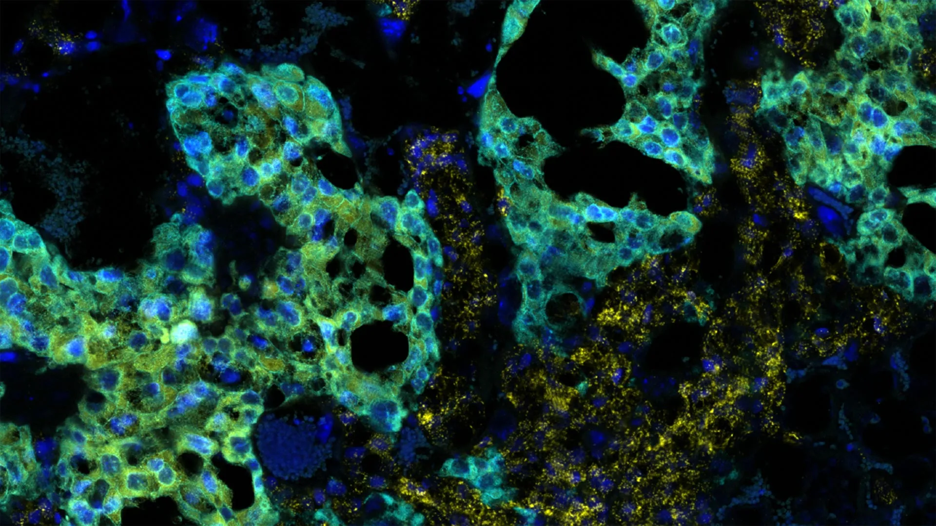
In their investigation, Cheadle and Sanchez Martin looked closely at why certain fetuses succumbed to developmental disruptions while others appeared entirely insulated from the effects of maternal inflammation. Their microscopic examinations zeroed in on a specialized population of cells known as spongiotrophoblasts. These cells occupy a critical spatial niche within the mouse placenta where maternal tissues and fetal cells intimately interact, forming the structural foundation for physiological exchange.
The imaging and histological analyses revealed a stark dichotomy. In fetuses that proceeded through gestation normally, the spongiotrophoblast layer remained structurally intact and healthy. Conversely, male fetuses that subsequently exhibited autism-like developmental traits displayed clear, localized cellular damage within this exact placental compartment.
From an immunological perspective, spongiotrophoblasts fulfill a paramount function: they facilitate maternal immune tolerance toward the developing fetus. Because an embryo contains genetic material inherited from both the mother and the father, it is technically a semi-allograft—foreign tissue that a fully functional immune system would ordinarily reject. The placenta maintains a delicate immunological ceasefire, shielding the fetus from maternal immune surveillance.
The CSHL findings demonstrate that maternal immune activation can severely disrupt this protective mechanism. When inflammation compromises the integrity of spongiotrophoblasts, the delicate immunological barrier breaks down. Consequently, the mother’s heightened immune response spills over into the fetal compartment, inadvertently subjecting the delicate, rapidly developing embryonic systems to inflammatory cytokines and signaling molecules that derail typical neurological and physiological maturation.
The Sexual Dimorphism Mystery: Why Males Bear the Brunt
One of the most persistent epidemiological puzzles in clinical neurology is the pronounced gender bias observed in autism spectrum diagnoses. Statistically, boys are diagnosed with ASD approximately four times more frequently than girls. Despite decades of epidemiological and clinical research, the biological underpinnings of this disparity have remained elusive, frequently attributed to a combination of diagnostic biases and complex genetic risk profiles.
The CSHL study injects a profound new biological variable into this conversation by uncovering a striking sex-specific divergence in embryonic response. Across every iteration of the experiment, autism-like traits manifested exclusively in male mice, whereas female littermates continued along a normative developmental trajectory, apparently shielded from the inflammatory insult.
What elevates this observation from an interesting curiosity to a major scientific breakthrough is the precise developmental timing involved. The sexual divergence manifested at day 12.5 of gestation—an epoch that precedes the major hormonal surges and the physical sexual differentiation of the brain and other anatomical structures.
"It’s really fascinating, because we’re looking at an age which precedes hormonal surges," Cheadle emphasizes. "It precedes sexual differentiation in the brain and many other regions. So, it’s surprising there would be a sex-based difference this early."
This temporal paradox suggests that the observed resilience in females and vulnerability in males operates independently of traditional sex hormones like testosterone and estrogen, which typically drive sexual dimorphism later in embryonic life. Instead, researchers hypothesize that fundamental genetic or cellular differences tied to chromosomal sex—such as the expression of genes on the X and Y chromosomes that escape X-inactivation or differential baseline cellular responses to oxidative stress—may confer an early, intrinsic protective advantage to female embryos.
Comparative Biological Frameworks in Neurodevelopmental Research
| Research Dimension | Traditional Focus | CSHL Study Approach | Key Implication |
|---|---|---|---|
| Primary Anatomical Focus | Fetal and Postnatal Brain | Maternal-Fetal Interface & Placenta | Shifts origin timeline and targets non-neural tissues. |
| Trigger Mechanism | Genetic Mutations & Idiopathic Factors | Maternal Immune Activation (MIA) | Highlights environmental contributions to developmental risks. |
| Chronological Window | Late Gestational & Postnatal Periods | Early Gestation (Day 12.5 in Murine Models) | Identifies an earlier critical period for structural vulnerability. |
| Sex-Based Outcomes | Generalized or Uncontrolled | Stark Dimorphism (Male Vulnerability vs. Female Resilience) | Provides a biological model for human diagnostic gender ratios. |
Implications for Future Research and Therapeutic Innovation
While the empirical data was generated within a murine model and cannot be directly translated as a definitive causative mechanism for human autism without extensive clinical corroboration, the implications for the broader scientific community are profound. The research fundamentally challenges the insular paradigm that neurodevelopmental disorders must be studied exclusively through the lens of neurology.
By framing the placenta as an active participant in neurodevelopmental outcomes, the CSHL findings suggest that therapeutic strategies might one day begin during pregnancy. If maternal immune activation and subsequent placental distress serve as a primary gateway for certain presentations of autism, then preserving placental integrity or modulating the maternal immune response during critical gestational windows could represent a viable avenue for prophylactic intervention.
Furthermore, decoding the natural protective mechanisms that allowed female fetuses to withstand the inflammatory assault in Cheadle’s experiments offers a blueprint for potential treatments. If scientists can successfully identify the biochemical pathways responsible for female embryonic resilience, those pathways could theoretically be harnessed, pharmacologically mimicked, or stimulated to protect vulnerable male fetuses or individuals facing high-risk inflammatory environments.
As Cheadle and his research team look toward the horizon, their experimental roadmap involves a granular, comparative analysis of both male and female embryonic tissue responses. By isolating the exact molecular signaling cascades that diverge following maternal immune activation, the researchers hope to map out the distinct developmental trajectories of both sexes.
Ultimately, this line of inquiry transforms our understanding of autism’s complex etiology. It bridges the gap between immunology, obstetrics, and neurology, underscoring the reality that the earliest chapters of neurodevelopment are written not by the brain alone, but through a complex, systemic dialogue between a mother, her placenta, and the changing biological environment of the developing child.

